Porous media, specifically open-pored asphalt concrete, is analysed with an (µ)XRCT (micro X-ray Computed Tomography) scanner. The scanner is a modular hardware system to implement advanced in situ setups
Object of Research and Objective
Porous media analysis, e.g. to detect micro fractures or investigate fatigue behaviour.
Procedure
The whole process of XRCT Scan
Sample preparation and parameters: Samples are prepared, mounted and positioned; X-ray source parameters and detector parameters are set
XRCT Scan: Recording of the primary / raw data (in this specific case the "radiograms") (in two phases: open beam (flat field) and dark images (dark field) (before the actual scan) and object projection image (the actual scan)
A radiogram, the output of step 2
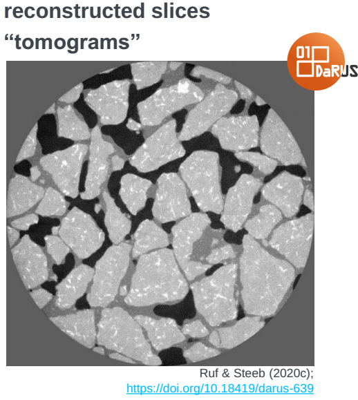
A tomogram, output of the image reconstructions ins step 3Image Processing/Reconstruction, that generates the research data (in this specific case "tomograms") from the primary/raw data. Core parts are the reconstruction algorithm, filters, linearization and normalization and correction methods
Post Processing, such as Segmentation, Quantification, Statistics
Data Curation, including Metadata Preparation and Repository ingest of the data
The primary research / raw data after the XRCT scan (this output serves as input for the subsequent step, or other research endeavours)
The research data generated by reconstruction algorithms, filters, linearization, normalization and correction methods from the primary/raw data.
Model
Discretization
Time: -
Space 2D Voxel size , $y$, ($z$), .., ROI off-size $x$, $y$, ($z$); Position $x$, $y$, ($z$)
Variables
Name
Unit
Symbol
dependent (measured) / independent (controlled)
Parameter
Name
Unit
Symbol
Process Informationen
Process Steps
Name
Description
Input
Output
Method
Parameter
Environment
Mathematical Area
Sample preparation and parameters
a. Samples are prepared, mounted and positioned b. X-ray source parameters and detector parameters are set
X-Ray and Detector parameters
Pre-Filters
XRCT Scan to generate the primary / raw data (in this specific case the "radiograms")
Recording of the data in two phases: a. open beam (flat field) and dark images (dark field) (before the actual scan) b. object projection image (the actual scan)
Sample; X-Ray and Detector parameters; Geometric Information
Projections, flat field images, dark field images
XRCT Scan
X-ray source parameters (tube voltage, tube flux, phys. pre-filters), detector parameters (binning mode, exposure time, number of image per projection, number of projection angles)
Image Processing/Reconstruction to generate research data (in this specific case "tomograms")
Image Processing/Reconstruction in following steps: a. Normalization and Linearization of the projection images -> Transforming the data into sinograms b. Correction of geometrical system misalignments c. Filters d. Reconstruction Algorithm e. Evaluation of the reconstructed data set
X-Ray and Detector parameters; Projections, flat field images, dark field images
(Reconstruction) Software performing the tomographic reconstruction based on the projection images as well as beam profile and dark images; Remark: Software was discontinued 2019
(Reconstruction) Software performing the tomographic reconstruction based on the projection images as well as beam profile and dark images, but next to analytical also algebraic reconstruction methods